
Ureteral Stent Placement/Removal
Department: Urology
Estimated Cost : $1000 - $3000
No Hospitals Available for this Treatment
Unfortunately, there are no hospitals currently offering this treatment. Please check back later for updates.
No Doctors Available for this Treatment
Unfortunately, there are no doctors currently offering this treatment. Please check back later for updates.
Ureteral Stent Placement and Removal
What Is a Ureteral Stent?
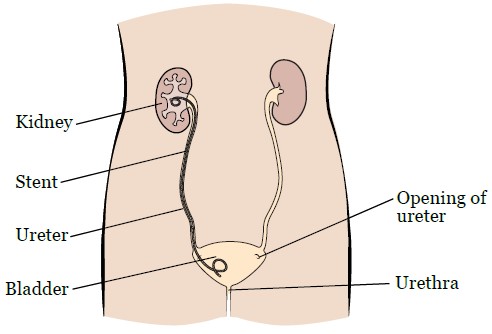
A ureteral stent is a thin, flexible tube inserted into the ureter — the duct that carries urine from the kidney to the bladder. The stent helps keep the ureter open and allows urine to drain freely when there is a blockage or narrowing due to medical conditions or after surgical procedures like pyeloplasty or stone removal.
Stents are often made of soft plastic and may have curled ends (called “double-J” or “JJ stents”) to keep them securely in place in the kidney and bladder.
Why Is a Ureteral Stent Placed?
Ureteral stents are used for a variety of reasons, including:
1. After Surgery:
- Following procedures like pyeloplasty, ureteroscopy, or kidney stone removal.
- To ensure the ureter stays open during healing and allows proper urine flow.
2. To Relieve a Blockage:
- Due to kidney stones, tumors, scar tissue, or inflammation that obstruct urine flow.
3. In Case of Ureteral Injury:
- After trauma or accidental surgical injury to the ureter.
4. Cancer-Related Obstructions:
- In cancers of the bladder, prostate, cervix, or other nearby organs compressing the ureter.
How Is a Ureteral Stent Placed?
Preparation:
- Typically done under general or spinal anesthesia.
- You may need to fast for 6–8 hours before the procedure.
- Blood and urine tests may be performed.
Procedure Steps:
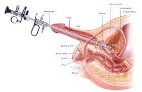
- A cystoscope (a thin tube with a camera) is inserted through the urethra into the bladder.
- The doctor visualizes the ureteral opening and inserts a guidewire into the ureter.
- The stent is threaded over the guidewire into the ureter and positioned properly.
- Once in place, the stent allows urine to bypass the obstruction and flow into the bladder.
- No external incision is usually required unless combined with another surgery.
 Get an Opinion